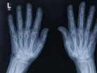
New Zealand scientists undertake the first 3D colour x-ray

The use of x-ray’s in the detection and diagnosis of potential fractures has remained relatively unchanged since its development in the 19th century. However, New Zealand scientists are set to revolutionise traditional black and white imaging through the use of particle-tracking technology.
From developing the Higgs Boston particle, scientists at CERN, the European Organization for Nuclear Research have utilised the technology used for the organisation’s Large Hadron Collider, CERN have successfully scanned the human body in 3D colour.
Similar to a camera, the device, named Medipix3, collects individual sub-atomic particles as they collide with pixels, producing high resolution, colourful images, guaranteeing a more accurate detection and diagnosis, not only with bone matter, but with surrounding tissues.
See also
- Pfizer is set to overhaul its overall operating healthcare model
- Research on the benefits of 3D printing in a trauma hospital
- Top 10 innovative healthcare pioneers
“This technology sets the machine apart diagnostically because its small pixels and accurate energy resolution mean that this new imaging tool is able to get images that no other imaging tool can achieve,” explained Professor Phil Butler, who developed the technology alongside his son, Anthony Butler.
Their company MARS Bioimaging Ltd are set to commercialise the 3D scanner and is linked to the University of Otago and Canterbury.
Through Medipix3, the colours generated will highlight the alternating energy levels and components, highlighting fat, water, calcium and potential disease markers, the duo have stated.
“In all current studies, promising early results suggest that when spectral imaging is routinely used in clinics it will enable more accurate diagnosis and personalisation of treatment,” Professor Butler added.
The technology is now set to be utilised in a new clinical trial for orthopaedic and rheumatology patients in New Zealand.
- How Zipline Uses Drones to Deliver Medicine Across AfricaTechnology & AI
- How is Schneider Electric Making the NHS More Accessible?Hospitals
- How DeepHealth is Using AI to Screen for Breast CancerTechnology & AI
- Martin Carpenter: How Tech is Reshaping Healthcare on JerseyDigital Healthcare


