
GE HealthCare Scales Photonova Spectra CT Research Network
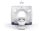
GE HealthCare has extended its clinical and research network to seven sites across North America, Europe and Asia in order to validate the Photonova Spectra, its next-generation photon-counting CT system.
After securing the requisite regulatory approvals, the programme is now moving into clinical use with Deep Silicon detector technology.
These multi-continent trials focus on key operational objectives for Healthcare Sustainability Directors and operational leads: reducing radiation dose, streamlining workflows into single scans, and improving diagnostic efficiency in high-volume care.

Photonova Spectra uses Deep Silicon detector technology to deliver ultra-high-definition spectral imaging in a single workflow.
By combining high spatial resolution with material and tissue characterisation, you can get quantitative data and spectral biomarkers from a single scan. That reduces the need for repeat scans, increases clinical throughput and reduces patient radiation dose in the more complex cases.
“Deep Silicon photon-counting CT has the potential to change not only what clinicians can see, but what they can understand from every scan – creating new possibilities for early detection, personalized care and diagnostic confidence,” says Chad Rowland, Executive Director for Global CT at GE HealthCare.
“Realising that potential requires close collaboration with the research and clinical community.
“By working alongside leading healthcare institutions around the world, we’re generating the clinical evidence and real-world experience needed to help define the future of photon-counting CT and ultimately improve care for patients.”
“Deep Silicon photon-counting CT has the potential to change not only what clinicians can see, but what they can understand from every scan ”
Clinical partners driving dose efficiency and workflow integration
Seven academic medical centres and specialty hospitals across three continents are currently evaluating image quality, spectral performance and workflow utility via formal research agreements.
- Keio University Hospital in Japan is evaluating quantitative and spectral imaging techniques, as well as emerging diagnostic applications, to define future clinical and research use cases.
- Italy’s Centro Cardiologico Monzino (IRCCS) evaluates cardiovascular imaging capabilities, focusing on quantitative assessment, tissue characterisation and cardiac workflow.
- Universitätsspital Zürich’s Department of Nuclear Medicine is assessing CCTA to optimise radiation dose levels, maintain diagnostic performance and refine clinical protocols.
- University of Wisconsin-Madison is building on previous system studies to assess quantitative performance, technical image quality metrics and standardised imaging protocols.
- Stanford Medicine in California is undertaking technical and human subject studies to evaluate image reconstruction techniques, presentation workflows, ultra-high-resolution structural visualisation and imaging biomarkers.
- UZ Brussel is evaluating the cardiac imaging capabilities, including coronary imaging and functional assessment, to optimise protocols for routine cardiovascular care.
- Rigshospitalet in Denmark is looking at a range of diagnostic applications, with a particular focus on paediatric and low-dose protocols, tissue characterisation and the use of spectral data in difficult cases.
Advancing low-dose protocols and cardiac diagnostics
Reducing ionising radiation exposure without compromising diagnostic accuracy is a major priority for modern healthcare organisations.
At Universitätsspital Zürich, the nuclear medicine team is developing dose-optimised protocols for coronary CT angiography, and at Rigshospitalet in Copenhagen, clinical teams are exploring paediatric and low-dose imaging to improve patient safety and radiation stewardship in these groups.
“Photon-counting CT offers exciting opportunities to advance coronary imaging by combining high spatial resolution with spectral information,” says Professor Philipp Kaufmann, Director of the Department of Nuclear Medicine at Universitätsspital Zürich.
“We aim to determine how best to translate these capabilities into robust, dose-efficient CCTA protocols that support confident clinical decision-making.”
Cardiac diagnostics are also a major focus at Centro Cardiologico Monzino (IRCCS) in Milan, where the focus is on how improved tissue characterisation and more efficient cardiac workflows can support clinical decision-making.
“Cardiovascular disease remains one of the world’s leading health challenges, underscoring the need for imaging technologies that enable more informed clinical decision-making,” says Professor Gianluca Pontone, Deputy Scientific Director; Director of the Department of Perioperative Cardiology and Cardiovascular Imaging; and Co-Director of the Sport Cardiology Unit, Centro Cardiologico Monzino IRCCS.
“We’re excited to work alongside GE HealthCare to better understand how Photonova Spectra can generate new clinical insights and help support more personalised cardiovascular care.”
Translational engineering and continuous product evolution
In addition to clinical validation, the seven-institution network provides a direct feedback loop between hospital clinicians, medical physicists and GE HealthCare engineering teams.
At Stanford Medicine and the University of Wisconsin–Madison, technical studies on image reconstruction and quantitative performance metrics are carried out, which in turn provide data to support standardisation of scanner performance across different clinical environments.
“Our collaboration with GE HealthCare has given our researchers an opportunity to explore how photon-counting CT can be applied across a range of technical and clinical questions,” says Adam Wang, PhD, Assistant Professor, Department of Radiology and, by courtesy, Electrical Engineering, Stanford University.
“We’re excited to continue our work with Photonova Spectra, building on our experience in reconstruction, image analysis and human-subject research to further investigate the capabilities of photon-counting CT and where it may create new opportunities for clinical practice and scientific discovery.”
During the trials, GE HealthCare engineers, scientists, and product teams are working hand in hand with hospital researchers to turn real-world clinical evidence into future hardware and software enhancements.
They're collecting protocol updates, dose reduction targets and workflow feedback from North America, Europe and Asia to inform ongoing product updates to the Photonova Spectra in healthcare systems around the world.
Company portals


Executives
Chad Rowland
Executive Director, Global CT