Scientists create first ever 3D cancer protein image
Scientists from Cancer Research UK have succeeded in creating the first ever 3D image of a protein that can help to prevent cancer.
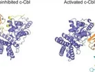
The team from the charity’s Glasgow-based facility, the Beatson Institute for Cancer Research, were able to map the structure of the c-Cbl protein, which controls cell growth.
Using high-tech X-Ray analysis the researchers found that when the c-Cbl protein is working as it should it changes shape.
This are now hoping this new discovery could lead to improved cancer treatments and prevention techniques.
To read the latest edition of Healthcare Global, click here
- Nanoparticles can boost the body’s response to vaccines
- Cholesterol-lowering statins can help to treat cancer
- The G-spot in women might not exist, say scientists
When the c-Cbl protein is defective or faulty, which it is known to be in some leukaemia patients, their cell growth is unregulated meaning cells divide excessively and could trigger cancer.
However, it was revealed that when the c-Cbl protein is switched on, it labels a cell receptor molecule for destruction.
In healthy cells the receptor amplifies a chain of cell signals resulting in normal cell growth, but in cancer cells these signals do not get switched off leading to uncontrolled cell growth.
By labelling the receptor molecule for destruction, the cell growth signal is switched off at the right time.
If c-Cbl cannot change to its active shape, it cannot label the receptor for destruction.
Dr Danny Huang from Cancer Research UK’s Beatson Institute in Glasgow was the lead author of the research, which has now been published in the journal Nature Structural & Molecular Biology.
Commenting on the findings, he said: “Using cutting-edge research techniques we’ve created the first 3D image of the structure of this protein, which is pretty incredible because in real life it’s about the size of a millionth of a hair width.
“We were intrigued to see that this protein actually changes shape when it’s switched on.
“Understanding the structure of this protein is vital because if the protein can’t be switched on it is more likely to cause cancer,” he added.
“So cracking the 3D structure is a step towards designing the cancer drugs of the future.”
Meanwhile, Dr Julie Sharp, Cancer Research UK’s senior science information manager, added: “Thanks to the generosity of the public, we’re able to fund a broad range of research projects like this across the UK to help us better understand how cancer cells grow, survive and spread.
“We hope these intriguing 3D structures of a key cancer protection protein will help pave the way to new approaches to tackle this disease more effectively,” she said.
The Healthcare Global magazine is now available on the iPad. Click here to download it.
Image c/o BBC News